Nervous system of a person from A to Z: Structure and Functions. Budova and significance of the nervous system Characteristics of the life and functions of the human nervous system
Nervous system control of the activity of all systems and organs and ensuring the communication between the body and the outer environment.
Budova nervous system
Structural unit of the nervous system is the neuron - the nerve clitina of the adolescents. In the whole of the nervous system, there is a cluster of neurons, constantly contacting each other with one of the additional special mechanisms - synapses. Behind the functions and structure, the following types of neurons are distinguished:
- Sensitive chi receptor;
- Efficiently - damaged neurons, as a direct impulse to higher organs (effectors);
- Zamikalni chi insert (conductor).
Mentally, the human nervous system can be divided into two great variables - somatic (or animal) and vegetative (or autonomous). The somatic system is important for the connection to the body from the outer middle, safe movement, sensitivity and fastness of the skeletal muscles. The vegetative system incorporates the processes of growth (breathing, exchange of speech, seeing and others.). Offenses to the system may be even more closely intertwined, only the autonomic nervous system of self-sufficiency and, according to the will of the people, cannot lie. The very same thing is also called autonomous. The autonomous system is divided into cute and parasympathetic.
The entire nervous system is composed of central and peripheral. The spinal and cephalic cord extend to the central part, and the peripheral system is a nerve fiber that enters the cephalic and spinal cord. As if marveling at the brain at the rose, it is clear that the wine is made up of white and orphan speech.
Sira speech - tse clusters of nerve clitins (with cob vіddіl vіdrostkіv, yakі vіd їkh іl). Okremі groupi syroї rechovina are called nuclei.
The white speech is made up of nerve fibers, covered with a myelin sheath (growths of nerve cells, from which the speech is made up). In the dorsal and cerebral cerebellum, the nerve fibers serve as pathways.
Peripheral nerves pod_lyayutsya on the rukhovi, sensitive and zmіshani, fallow, from which fibers the stench is formed (rukhovi chi sensible). The body of neurons, which are composed of sensitive nerves, rest at the nerve nodes in the brain. The bodies of the organ neurons are found in the organ nuclei of the brain and the anterior horns of the spinal cord.
Functions of the nervous system
Nervous system nadaє rezny infusion on the body. Three main functions of the nervous system - tse:
- Puskov, which calls out the function of the organ (secretion of the burrow, shortness of the m'yazi thinly.);
- Vasomotor, which allows you to change the width of the lumen of the vessels, regulating the flow of blood to the organ;
- Trophic, which reduces or increases the exchange of speeches, and also, slowing down the sourness of those living speeches. Tse allows you to gradually satisfy the functional camp of the organ that needs it in the sourness of the living speeches. If impulses are sent along the rukhovy fibers to the practical bony m'yaza, which call out її fast, then at once the impulses arrive, which boost the exchange of speech and expand the vessel, which allows you to secure the energy capacity.
Illness of the nervous system
Together with endocrine diseases, the nervous system plays a vital role in the functioning of the body. It is worthy of praise for the work of all systems and organs of the human body, as well as the spinal, cephalic and peripheral systems. The motor activity and the sensitivity of the body are stimulated by nervous endings. And the hearts of the vegetative system are inverted by the heart-vascular system and other organs.
Therefore, the disruption of the functions of the nervous system affects the work of all systems and organs.
Infection of the nervous system can be divided into infectious, recessive, sustin, traumatic and chronically progressive.
Falling ailments are genomic and chromosomal. The most common and widest chromosomal disease is Down's disease. These ailments are characterized by such signs: damage to the side of the musculoskeletal apparatus, endocrine system, lack of rosacea.
Traumatic damage to the nervous system is blamed on clogged masses and injuries, or when crushing the brain or spinal cord. Such ill-health, as a rule, is accompanied by vomiting, boredom, a waste of memory, a breakdown in information, and a waste of sensitivity.
Sudine diseases are most importantly developed on aphids, atherosclerosis and hypertensive disease. To tsієї category it is possible to include chronic cerebrovascular insufficiency, damage to cerebral blood flow. Characterized by onset symptoms: attack vomit and nudoti, head bіl, Damaged rukhovoї activity, change in sensitivity.
Chronically progressive ailments, as a rule, develop as a result of disruption of metabolic processes, infection, intoxication of the body, or through anomalies of the nervous system. Before such ailments, sclerosis, myasthenia gravis and others can be seen. The disease progresses step by step, reducing the incidence of certain systems and organs.
Causes of the disease of the nervous system:
Possibly placental way of transmission of ailments of the nervous system during pregnancy (cytomegalovirus, rubella), as well as peripheral system(Poliomyelitis, skaz, herpes, meningoencephalitis).
Crimson, the nervous system is negatively affected by endocrine, heart, nirk disease, indigestion, chemistries and medical care, important metal.
1. Budova and functions of the nervous system. Glia.
2. Reflex. Reflex arcs. Classification of reflexes.
3. Vikovi features of the brain and spinal cord.
1. Budova and functions of the nervous system. glia
The nervous system regulates and coordinates the activity of all organs of the prosecutor's office and systems, and improves the integrity of the functioning of the body. Zavdyaki їy zdіysnyuєtsya zv'yazok to an organism іz ovnіshnіm sredovischem that yoga adaptation to minds, scho constantly zmіnuyuetsya. Nervous system is the material basis of the life of a person, thoughts, behavior, movement.
The head and spinal cord can be seen to the central nervous system. Having offended the stench, it is evolutionary, morphologically and functionally related to each other and without a sharp boundary to pass one into one.
Functions of the nervous system
1. Protect the body from the outer environment.
2. Take care of the mutual interaction of all the elements in the body among themselves.
3. Ensure the regulation of trophic functions, tobto. regulation of speech exchange.
4. Nervous system, cerebral cortex, substrate of mental activity.
Functionally, the nervous system is divided into somatic and autonomous (vegetative), anatomically - into the central nervous system and the peripheral nervous system.
The somatic nervous system regulates the work of skeletal masses and ensures the sensitivity of the human body. Autonomous (vegetative) nervous system regulates the exchange of speech, the work of internal organs and smooth membranes.
The autonomic nervous system innervates the internal organs. It will also take care of the trophic innervation of the bones, other organs and tissues of the nervous system itself.
The peripheral nervous system is made up of numerous paired nerves, nerve plexuses and knots. Nerves deliver impulses from the central nervous system directly to the working organ - m'yaza - information from the periphery in the central nervous system.
The main elements of the nervous system are nerve cells (neurons). Confirmation of the cellular theory of the future nervous system was taken away by the help of electron microscopy, as it showed that the membrane of the nervous clitina guesses the main membrane of other clitinae. Vaughn appears to be a succulent stretching of the upper surface of the nervous clitinum and the cremation of the other clitinum. The skin nerve cell is an anatomical, genetic and metabolic unit, like cells and other tissues of the body. Nearly 100 billion nerve cells are located in the human nervous system. Shards of the dermal nerve cell are functionally connected with thousands of other neurons, the number of possible variants of such connections is close to infinite. Nervous clitin should be considered as one of the lines of organization of the nervous system, which acquires molecular, synaptic, subclitinal lines with supraclitinal lines of the canal neural networks, nerve centers and functional systems of the brain, which organize behavior.
Budov neuron. The body of the neuron, as if tied with sprouts, is the central part of the neuron and ensures eating of other parts of the clitin. Open the body with a spherical membrane, as it is two balls of lipids with a parallel orientation, which make up the matrix, which avenges proteins. The body of the neuron is the nucleus of the nucleus, which avenges the genetic material.
The nucleus regulates the synthesis of proteins in all cells and controls the differentiation of young nerve cells. The cytoplasm of the body of the neuron has a large number of ribosomes. Some ribosomes roam freely in the cytoplasm, one by one, or they create a clump. Other ribosomes are attached to the endoplasmic reticulum, which represents the internal system of membranes, tubules, and puffers. Ribosomes are attached to the membranes to synthesize proteins, which are then transported from cells. Accumulation of the endoplasmic reticulum with budding into the new ribosomes becomes characteristic of neuron neuron illumination - Nissl's substance. Heaps of smooth endoplasmic reticulum, which do not contain ribosomes, form part of the Golgi apparatus; It is suggested that it may be important for the secretion of neurotransmitters and neuromodulators. Lysosomes are arranged in membranes for accumulation of various hydrolytic enzymes. Important organelles of nerve cells are mitochondria - the main structures of energy production. On the inner membrane of mitochondria, all enzymes of the cycle are located citric acid- the most important route of the aerobic pathway for the breakdown of glucose, which is dozens of times more effective for the anaerobic pathway. Nervous cells also have microtubules, neurofilaments and microfilaments, which vary in diameter. Microtubules (diameter 300 nm) go from the body of the nerve cell to the axon and dendrites and the internal transport system. Neurofilaments (diameter 100 nm) grow only in nerve cells, especially in the great axons, and form part of the transport system. Microfilaments (diameter 50 nm) are well manifested in the growths of nerve cells, which grow, stinks take the fate of some types of interneuronal diseases.
Dendrites are wood-like-gills of a neuron's growth, its head receptive field, which ensures the collection of information, like coming through the synapse from other neurons, or directly from the middle. When the body is farther away, dendritic discoloration occurs: the number of dendritic needles increases, and their diameter sounds. On the surface of the dendrites of rich neurons (pyramid neurons of measles, cells of Purkin's cerebellum and in) there are spines. Spike apparatus є warehouse tubular systems in the dendrite: microtubules, neurofilaments, Golgi apparatus and ribosomes are located in the dendrites. Functional maturation and the cob of active activity of nerve cells develops with the appearance of spines; Trivale attaching the necessary information to the neuron leading to the dismantling of the spines. The appearance of spines increases the adhesion to the surface of the dendrites.
The axon is a single, sounding long-term excitatory neuron, which serves for a quick wake-up call. In the province of wines, it can grow to a great extent (up to 1000) number of small beetles.
Nervous clitiny vikonuyut low zagalnyh functions, contributions to the support of the power processes of the organization. The price of the exchange of speeches with a navkolyshnim middle, the conversion of that vitrachannya energy, the synthesis of proteins and in. In addition, nerve cells win power only to them specific functions to enable them to process, process and collect information. The neurons of the building accept information, reshape (code) її, quickly transmit information in specific ways, organize interaction between the Cosmos and other nerve cells, save and generate information її. For the purpose of understanding these functions, the neurons can be polar organization with a subdivision of inputs and outputs and a number of structural and functional parts.
Classification of neurons. Neurons are divided into the following groups: after the mediator, which is seen in the ends of axons, they are divided into adrenergic neurons, cholinergic, serotonergic, etc.
The neurons of the somatic and vegetative nervous systems are seen in the fallow in the CNS.
In direct information, the following neurons are divided:
Afferents that receive additional receptors for information about the outer and inner middle of the body and transmit it to the central nervous system;
Efferent, which transmit information to working organs - effectors (nervous cells, which innervate effectors, are sometimes called effectors);
Inserts (interneurons), which ensure the interaction between CNS neurons.
By the injection, you can see neurons chirping and chirping. According to their activity, background-active and “momentary” neurons are distinguished, which are less likely to be stimulated in the course of development. The background-active neurons are charged with a bright little pulse generation, the fragments of some neurons are discharged without interruption (rhythmically and arrhythmically), others - in bursts of pulses. The interval between pulses at the patch becomes milliseconds, between bursts - seconds. Background-active neurons play an important role in improving the tone of the central nervous system and especially measles.
According to the sensory information that is received, neurons are divided into mono- and bipoly-sensors. Monosensory - neurons to the center of hearing in the cerebral cortex. Bisensor neurons are heard in the secondary zones of the analyzers in the cortex (neurons of the secondary zone of the analyzer in the cortex of the great brain react to light and sound signals). Polysensory neurons - ce neurons of the associative zones of the brain, motor measles; stench reacts to teasing receptors of the skin, eyes, auditory and other analyzers.
Nerve cells are tied together by numerical connections: the end of the axon cleavage of one neuron sticks to the dendrite of the next neuron, and the axon cleavage wraps around the entire body of the other neuron. The place of a close dotic of neurons is called a synapse.
Synapse is a structural solution that ensures the transfer of excitation from the nerve cell to the nerve cell or from the nerve cell to the cell of the working organ. The term “Synaps” was propagated by the English physiologist C. Sherrington.
Whether a synapse is made up of 3 parts - a presynaptic fiber, a synaptic gap and a postsynaptic fiber.
The presynaptic part is formed from the terminal part of the axon covered by the presynaptic membrane. In the middle are bulbs - vesicles, which avenge chemical speech - a mediator.
The synaptic gap is filled with native, close to blood plasma.
The postsynaptic function of the representations of the postsynaptic membrane, where there are chemoreceptors that are sensitive to the singing mediators.
The synapse has a large number of mitochondria.
The electrical impulse of the awakening, similar to the axon, reaches the synaptic bulbs, as a result, it wakes up and rises. From the bulb, acetylcholine comes out, which through the pores of the presynaptic membrane is located at the synaptic gap and enters into a chemical interaction with the receptors of the postsynaptic membrane. As a result, rux cations are attached to potassium and significantly increase rux cations to sodium, stench collapses in the middle of the nerve fiber and on the surface of the postsynaptic membrane, a negative charge is generated - depolarization occurs. At a glance, the awakening of the veins is transmitted to the outer nerve cell.
Neuroglia, otherwise glia, was first seen as a group of elements of the nervous system in 1871. R. Virkhovim. Cells of neuroglia cover the space between neurons, making up 40% of the brain. Over the course of the century, the number of neurons in the human brain changes, and the number of glial cells increases. Beyond the size of the glial cells, 3-4 times less than the nerve cells, their number of magnitudes increases with age (the number of neurons changes). The thila of neurons, like that of yogo axons, is sharpened by glial clitins. Glial cells perform a few functions: supporting, suppressing, isolating, exchange (the supply of neurons with living speeches). Microglial cells start to phagocytosis, rhythmic changes in their contraction (the period of rapidity - 1.5 minutes, relaxation - 4 minutes). The cycles of change are obligatory repeated through the skin 2 - 20 years. It is important to note that the pulsation joins the protrusion of the axoplasm in neurons and flows into the struma of the intercellular nerve. The process of destruction in
neurons and electrical phenomena in glial clitins, perhaps, interact.
Glіya vykonuє such functions:
Ensure the normal functioning of the neurons and the whole brain;
Ensures superficial electrical isolation of the bodies of neurons, their progeny, synapses to turn off inadequate interplay between neurons in case of extensive stimulation of trophic functions by neuron lances in the brain.
2. Reflex. Reflex arcs. Classification of reflexes
At the heart of the activity of the nervous system lies a reflex character, that is, a reflex.
A reflex is a reaction in the organism, which is blamed on various subdivisions of the outer or inner environment and is dependent on the help of the central nervous system.
In the 17th century, R. Descartes saw miraculous changes in the group of victorious children, which are blamed afterward on the nervous system of subdivisions, like they are injected into the body. They turn out to look like end reactions in the mind.
The anatomical path, which creates a reflex, is called a reflex arc (Fig. 5.3). Won maє 5 lanok:
1) receptor - illumine, which is irritating
2) afferent or sensory, sensitive, pre-central pathway
3) nerve center - a branch of the central nervous system
4) efferent, or ruhovy, motor vіdtsentrovy way
5) working body chi efector
The reflex is not based on a linear scheme, but on the type of reflex ring (for Anokhin). Dodaetsya shosta lanka - zvorotny afferent call.
The attachments of the links secure the nerve centers of information to the working organ and give the possibility to make the necessary corrections in the formation of the reflex act.
Reflex arcs can be different for folding:
Monosynaptic (two neurons);
Polysynaptic (3 and more neurons).
3. Vikovi features of the brain and spinal cord
In a newborn, the spinal cord should be 14 cm at the age of 14 cm, up to two years - 20 cm, up to 10 years - 29 cm. two sweats are well expressed, and the central channel is wider, lower in a grown-up one. In the first two years, it is necessary to change the enlightenment of the central channel. The volume of the white speech is growing faster, the lower volume is the gray speech.
Sensitivity is important for the life of the organism. For the help of sensitivity (obviousness), a link is established to the body from the outermost center and orientation to the new. Sensitivity to look at a glance about analyzers.
The analyzer is a foldable nerve mechanism, which spriymaє razdratuvannya, carry out yoga in the brain and analyze it, to spread it on the surrounding elements. The analyzer of maє roztashovaniya on the periphery priymaє provіdnikovy apparatus (nervі vіdniki) and the central apparatus, which is found in the cortex of the brain. The cortical product of the analyzer creates analysis and synthesis of various teasings of the outer world and the internal environment of the organism. Distinguish between good, hearing, smelling, savory and skinny analyzers.
The peripheral apparatus of the analyzer is called the receptor. Receptors receive razdratuvannya and reshape them in the nerve impulse. Exteroreceptors, which receive teasing from the inner middle, interoreceptors, which receive teasing from the internal organs of the body, and proprioreceptors, which receive teasing from the m'yaziv, tendon, and joint. The impulses in the proprioceptors blame the tendon against the tension of the tendon, m'yazіv and orient the body in the right position of the body in the open air. Seeing the sensitivity of bindings with the type of receptors. Bolova, temperature and tactile sensitivity is associated with exteroreceptors and is carried to the surface sensitivity.
It’s almost like the position of the toulub and the kіntsіvok in the open space (m'yazovo-suglobov it feels like), vіdchutta the grip of that vaga, the vibratіyna sensitivity po'yazanі z proprioceptors that can be seen to deep sensitivity. Distinguish the same folding and sensitiveness: a little bit of localization of teasing, stereognosis (recognition of objects on dotik) and others.
The most important connection of the nervous system with the normal vital functions of the body reaches the heart of the fact that different organs, parts of the body and whole physiological systems are not designed at the same nerve centers. So, for example, in the sensitive zones of the measles of the great pіvkul є special dilyanki, where sensitive impulses are projected from the legs, the coat, the hands, the guise. This principle of somatotopic projection (projection of parts of the body) is common in the brains of rich children. On the level of the spinal cord, the somatotopic projection has its own form: parts of the body are presented segment by segment. The central segments schematically look like transverse swags on the tulub, the later ones - on the tips, and the concentric stakes - on the faces. The skin segment of the body corresponds to the segment of the spinal cord.
In the functioning of the nervous system, signs of hierarchy are observed: that function itself is forward regulated by the lower centers, from which things are awakened. Such a rich surface of regulation significantly promotes the superficiality of the work of the nervous system and at one and the same time exudes an evolutionary history.
Century features of the brain.
Masa of the brain of a newborn to become an average one 390 r. Until the end of the first fate of life, it will be won, and up to 3-4 years - it will be built. After 7 years of age, the mass grows more and more and the maximum value reaches up to 20-29 years (1355 g - for men and 1220 g - for women). Until about 60 years of age, the brain does not change, and after 60 years, a day of change is indicated.
At the time of birth, most of the nuclei of the stovbur were well decomposed, and the children of their neurons were myelinated. The structure of the middle brain is insufficient for differentiation. Such nuclei, like a red kernel, black speech, ripen in the postnatal period, forming the lowest passageways of the extrapyramidal system. The middle brain of the new people's branch is clearly good. At the time of birth, the differentiation of specific and non-specific nuclei of the thalamus, which is why all the sensitivity is formed. Residual maturation of the thalamic nuclei will end in about 13 years. Until the 2nd-3rd age, most of the hypothalamic nuclei are already formed, but there is still a residual functional maturity until 15-16 years of age.
Intensive development of the structures of the cerebellum occurs during the period of state maturation. In a single child, the mass of the cerebellum becomes 90 g. Up to 7 years, the mass of the cerebellum is grown up (130 g).
ANATOMY AND PHYSIOLOGY OF THE CENTRAL NERVOUS SYSTEM.
VISCHA NERVOUS DIALNIST. SMART REFLEXI
2. Looked at the brain
2.1. Veliki pіvkuli (chastki, furrows, zvivini, sіra ta bіla)
speech)
2.2. Budov's stovbur brain (dovgasty brain, posterior brain, middle
2.3. Budov of the crotch (thalamus, epithalamus, metata-
lamus, hypothalamus)
2.4. Cerebral cortex
1. Spinal cord (topography and budova)
The spinal cord is more ancient than the central nervous system. The spinal cord appears to be a long, cylindrical shape, flattened from front to back with a narrow central canal in the middle.
The dozhina of the spinal cord has grown to an average of 43 cm, weight - close to 34-38 g, which becomes approximately 2% of the head of the brain.
The spinal cord is segmental. On the rivers of the great potile lapel, cross at the head brain, and on rivers 1 - 2 of the transverse ridges end with a cerebral cone, from which the terminal / end / thread enters, honed by the roots of the transverse and cranial spinal nerves. In the areas of the nerves exit to the upper and lower ends, there is sweating. Qi sweating is called shiny and cross / cross-krizhovim /. In the uterine development, there are no signs of sweating, the cervical sweating is less on the level of the V-VI cervical segments and the transverse-crown at the spheres of the III-IV transverse segments. There are no morphological inter-segments of the spinal cord, so I divided them into functional segments.
There are 31 pairs of spinal nerves entering the spinal cord: 8 pairs of cervical, 12 pairs of thoracic, 5 pairs of transverse, 5 pairs of cranial and a pair of cupric.
Inner Budova spinal cord
The spinal cord is composed of nerve cells and fibers of gray speech, which may look like the letter H or panicle on a transverse view. On the periphery of the gray speech, there is a white speech, filled with nerve fibers. At the center of the gray speech, the central canal is expanded to avenge the spinal cord. The upper ending of the channel is connected with the IV duct, and the lower ending is the ending duct. In the Syrian speech, the anterior, lateral and posterior horns are distinguished, and on the transverse view, the anterior, lateral, and posterior horns are distinct. In the anterior horns, there are ruffled neurons, in the posterior horns - sensitive neurons, and in the biceps - neurons that satisfy the centers of the sympathetic nervous system.
The human spinal cord contains about 13 neurons, of which 3% are motor neurons, and 97% are inserted. Functionally, spinal cord neurons can be divided into 4 main groups:
1) motoneurons, or rukhovi, - cells of the anterior horns, the axons of which form the anterior roots;
2) interneurons - neurons that take information from the spinal ganglia and from the posterior horns. Qi neurons respond to pain, temperature, tactile, vibrational, proprioceptive teasing;
3) sympathetic, parasympathetic neurons are more importantly developed in beetle horns. Axons of these neurons emerge from the spinal cord at the warehouse of the anterior roots;
4) associative cells - neurons of the upper apparatus of the spinal cord, which establish links in the middle and between segments.
In the middle zone of the gray speech (between the posterior and anterior horns) of the spinal cord, the intermediary nucleus (Kahal nucleus) with clitins, the axons of which go uphill or down by 1-2 segments, satisfying the border. It is similar to a tether on the top of the posterior horn of the spinal cord - this tether makes up the so-called dragline speech and viconizes the function of the reticular formation of the spinal cord.
Sira speech of the spinal cord establishes the segmental apparatus of the spinal cord. The main function of the development of congenital reflexes in the development of /internal externality/.
The white speech is subdivided into three funiculi from the skin side: anterior, posterior, and posterior.
White speech is filled with myelin fibers. Bundles of nerve fibers, which link the various branches of the nervous system, are called conduits of the spinal cord. You can see three types of guiding paths.
1. Fibers that support the spinal cords of the spinal cord in different equals.
2. Move / afferent, inferior / fibers that go from the brain to the spinal cord on the back of the anterior horns.
3. Sensitive / afferent, vishіdnі / fibers that direct to the centers of the great brain and cerebellum.
Usі vyskhіdnі kіrkovі ways are made up of three neurons.
The first neurons are scattered in the organs of the senses, ending in the spinal cord or in the stovburov part of the brain.
Other neurons are located in the nuclei of the spinal cord and the brain, and terminate in the nuclei of the thalamus and hypothalamus. The number of neurons is responsible for docentre vishіdnі paths.
The third neurons lie at the nuclei of the crotch / in the nuclei of the thalamus / for skin and m'yazovo-glom-bog sensitivity, for oral impulses in the narcissistic body, scent impulses in the sac-like bodies. Growths of the third neurons terminate on the clitins of the central Kirk centers /sounding, auditory, scenting and sensitiveness/.
In the middle of the central nerve pathways, it is necessary to see the Kirk-spinal cord /pyramids/ and the Kirk-cerebellar paths.
The function of the spinal cord is to act as a coordinating center for simple spinal reflexes /colon reflex/ and autonomic reflexes /shortening of the slash mihur/, as well as linking between the spinal nerves and the brain.
The spinal cord has two functions: reflex and conductor.
Reflex functions. Nerve cells in the body are connected with receptors and working organs. The rukhovi neurons of the brain are innervated by the mucosa of the tuba, kintsivok, shii and dichalna mucosa - the diaphragm and intercostal mucosa.
The reflex action of the spinal cord is controlled by segmental reflex arcs.
The conductor functions are counted for the rahunok of the upper and lower paths. Qi paths connect the main segments of the spinal cord one by one, as well as from the brain.
Bleeding of the spinal cord
The blood supply to the spinal cord is supplied by the spinal artery, deep cervical artery, intercostal, transverse, lateral cranial arteries.
Century features
In a newborn, the spinal cord should be 14 cm at the dovzhin, up to two years - 20 cm, up to 10 years - 29 cm. 19 gr. The new-born good has two sweats, and the central channel is wider, lower in the grown-up one. In the first two years, it is necessary to change the enlightenment of the central channel. The volume of the white speech is growing faster, the lower volume is the gray speech.
2. Looked at the brain
2.1. Veliki pіvkuli (chastki, zvivini, sіra ta bіla rechovina)
The cephalic brain is composed of: dovetail, posterior, middle, intermediate and terminal brain. The posterior brain is subdivided into the brain.
The cephalic brain is found near the empty cerebral skull. May swell the upper lateral surface and the lower surface - flattened - the basis of the brain
The mass of the brain has grown up people from 1100 to 2000 grams, from 20 to 60 years of age, the mass is subject to maximum and constant, after 60 years it changes slightly. No absolute, no visible mass of the brain is not an indication of the degree of rozum development. Mass of the brain of Turgenev 2012, Byron 2238, Cuve 1830, Schiller 1871, Mendelev 1579, Pavlov 1653 The brain stem is composed of neurons, nerve tracts and blood vessels. The head brain is made up of 3 parts: the pivkul of the great brain, the brain and the brain stovbur.
Pіvkuli great brain reach the maximum development in humans, yak vinikla pіznіshe for іnshі vіddіli.
The great brain is made up of two pivkul - right and left, as if they were tied one with one commissure / commissure / - callused body. The rights of that liva pivkulі to share for the help of the late spring. Under the commissary there is a crypt, which is two bent fibrous strands, yak in the middle part of the joint between themselves, and diverge in front and behind, satisfying the steps and the bottom of the crypt. In front of the stovpiv of the crypt there is an anterior commissure. Between the corpus callosum and the crypts, a thin vertical plate of brain tissue is stretched - a septum gap.
Pivkuli can be seen on the upper lateral, medial and lower surfaces. Upper lateral swell, medial - flat. Turned up to such a superficial inshoї pivkuli, and lower irregular shape. On three surfaces, there are deep and dry furrows, and between them zvivini. Borozny - burial ground between Zvivins. Zvivini - the rise of brain speech.
The surface of the pivkul of the great brain is reinforced with edges. The upper edge, the lower lateral edge and the lower vertical edge. In the expanse between two pivkuly enter the crescent of the great brain - the great crescent-shaped dross, which is a thin plate of hard tunic, as if penetrating into the later crevice of the great brain not reaching the corpus callosum and water-kremlin one in one right and left pivkul. The most protruding poles of pivkul took away the name of the poles: the frontal pole, the tilichny pole and the skronevy pole. The relief on the surface of the great brain's pivkul is more folded and connected with the presence of more less deep furrows of the great brain and ruffled between them, roller-like bottoms - the ringing of the great brain. Glybina, the length of some furrows and zvivin, their shape is directly narrower.
The skin pivkulya is divided into parts - frontal, tim'yana, potilichna, skronev, ostrivtseva. Central furrow / Roland's furrow / in the frontal part of the frontal part in the thym'yanoy, lateral furrow / Silvia's furrow / in the middle of the ridge in the frontal and thym'yanoї, thym'yano-potilichna distribution of the thym'yana and the potilichnu share. The lateral furrow is laid up to the 4th month of intrauterine development, the thyme-bearing furrow is the central one up to the 6th month. In the intrauterine period, gyrification is observed - molding zvivin. The three furrows are blamed for the first ones, and they are blasted with a great deep. Nezabar to the central furrow, another pair of parallel ones is added: one passes in front of the central one and is called in front of the central one, as it splits into two - upper and lower. The second furrow grows behind the central one and is called post-central.
The post-central furrow lies behind the central furrow and may be parallel to it. Between the central and post-central furrows there is a post-central zvivina. On the mountain, it passes to the medial surface of the pivculus of the great brain, degrades from the precentral fold of the frontal part, making the paracentral chasm from it at once. On the upper lateral surface of the pivculus, below, the postcentral sulcus also passes at the precentral sulcus, hoaring the central furrow below. Vaughn is parallel to the upper edge of the pivkul. Burn out in the inner parietal furrow, there is a group of small zvivins that took away the name of the upper thyme chasm. Below the furrow lie the lower thym'yana chasm, at the borders of which one can see two links: the upper edge and the lower one. Nadkrajova zvivina okholyuє the end of the lateral furrow, and the apex - the end of the upper cover furrow. The lower part of the lower thyme chasm and the lower parts of the post-central ring adjoined to it at the same time lower part in front of the central ring, which hang over the island's part, make up the frontal-thyme lining of the island.
Parts of the brain
The dorsal and lateral surface of the measles of the brain was taken to be divided into chotiri parts, so they took away the names of the external bones of the skull: frontal, tim'yana, tilichna, skroneva.
Potilichnaya part of the rostashovuetsya behind thyme-potilichnoi furrow and її mental prodovzhennia on the upper lateral surface pivkuli. The couples with other parts of the won may have small differences. At the back, a tiled section ends with a tiled pole. Furrows and ridges on the upper lateral surface of the politic area are even more variable. Most often and more often than not, there is a transverse sweaty furrow, which is not continued behind the inner parietal furrow of the thymus part of the brain.
Skronev's part is borrowed by the lower lateral vіddіl pіvkulі and vіdokremlyuієє vіd blovoї і іm'yanoї frequent deep lateral ї furrow. The edge of the skronevy part, which covers the ostrivtsev part, deleting the name of the skronevy lining of the island. The anterior part of the skeletal part satisfies the skronevy pole. Two furrows are visible on the side surface of the crown part, the upper and lower crown may be parallel to the lateral furrow. Zvivini skronevy part of the orientated vzdovzh borozen. The upper side of the ridge is cut between the lateral furrow at the top and the upper side at the bottom. On the upper surface of the ridge, tacked in the depth of the lateral furrow, 2-3 short ridges of the ridges (Heschl's ridges) are cut, separated by transverse ridge furrows. Between the upper and lower skronevy furrows there is a middle skronevy zvivina. The lower lateral edge of the skronevy section is occupied by the lower skroneval zvivina, which is surrounded by a furrow of the same name. The back end of the ring is continued in the utility room.
Above the corpus callosum, water-creaming from other branches of the pivkul, there is a furrow of the corpus callosum. Rolling back the corpus callosum, the furrow is straight down and forward and continues into the hippocampus furrow or hippocampal furrow. Above the furrow of the corpus callosum there is a belt furrow. The furrow begins to the front and bottom of the corpus callosum, rises uphill, then turns back and follows parallel to the corpus callosum furrow, ends more and behind the corpus callosum under the name of the dark furrow. On the level of the ridge of the corpus callosum, in the middle of the furrow, uphill, there is a regional part, which goes uphill and up to the upper edge of the pivkul of the great brain. Between the furrow of the corpus callosum and the belt furrow there is a belt furrow that hobbles the corpus callosum in front, the beast and behind. From behind and down, in the form of a corpus callosum, the lumbar ring sounds, satisfying the isthmus of the cingulate ring.
Between the furrow of the corpus callosum and the belt furrow there is a belt furrow that hobbles the corpus callosum in front, the beast and behind. From behind and down, in the form of a corpus callosum, the lumbar ring sounds, satisfying the isthmus of the cingulate ring.
Medial surface of pivculi. Mustache parts of the pivkul, krіm ostrivtsevoi, take part in the illumination of the medial surface.
On the medial surface of the tiling part, two deep furrows are dug out, which are angry one by one under the warm hood, opening backwards. This is a thyme-polylychna furrow, which strengthens the thyme part in the potilichny, and a spur furrow, which starts on the medial surface of the potilichny pole and straight ahead to the isthmus of the cingulate ring. A plot of a potilichny part, which lies between a thym'yano-potilichnaya and a spur furrow and has the shape of a trikutnik, with its top turned up to the point of angry furrows, is called a "wedge". Good mark on the medial surface of the pivkuli is the spur furrow between the animal tongue zvivina, which stretches from the potilichny pole from the back to the lower part of the isthmus of the belt zvivin. Bottom view of the pagan ring
collateral furrow, which lies even lower surface pivkuli.
The anterior ventilated the lower surface of the frontal part of the pivkul, behind which the skronevy pole protrudes, and there are also the lower surfaces of the skronevy and the tilichny chasm, which can pass one into the other without commemorated borders.
On the lower surface of the frontal part, which is lateral and parallel to the posterior fissure of the great brain, there is a sniff furrow. From the bottom to it lie the scent cibulina and the scent tract, which passes posteriorly into the scent tricot, in the area of \u200b\u200bwhich one can see the medial and lateral scent ducts. The dilyanka of the frontal section between the late crevice of the great brain and the scent furrow took away the name of the straight line. On the top of the frontal part, which lies lateral to the scent furrow, it is divided by shallow ophthalmic furrows into sprats that are variable in shape, roztashuvannyam and razmirami zvivin eyes.
At the back of the lower surface of the lower surface of the pivculus, a collateral furrow is clearly visible, which lies to the bottom and laterally in the lingual ring on the lower surface of the pelvic and skeletal folds, laterally in the view of the parahippocampal ring. Dekilka forward in front of the anterior end of the collateral furrow there is a nasal furrow, which surrounds the lateral side of the curvature of the end of the parahippocampal zvivini - a hook. Lateral to the collateral furrow lie medial to the tilice-skroneva zvivina.
Mіzh tsієyu zvivinoy and roztashovanoy nazvani vіd neї lateral potilichno-skronevy zvivinoy zvivinoy potilichno-skroneva furrow. It is not a furrow, but the lower lateral edge of the pivkul of the great brain, that serves as a cordon between the lateral tilichno-skronevy and lower skronevy zvivins.
The upper-lateral surface of the pivculi is the frontal part, which is located in the anterior part of the dermal pivculi of the great brain, which ends in front with the frontal pole and surrounds the lateral (sylvian) furrow below, and the deep central furrow in the back. A number of brain waves, rotting importantly on the medial surface of the pivculus and as a substrate for molding such hot beds, like sleeplessness, sleep, emotions and ing, are seen under the name "limbic system". Shards and reactions were formed in connection with the primary functions of the scent (in phylogeny), their morphological basis - they formed the brain, as they develop from the lower branches of the brain michur and lie down to the so-called scent brain. The limbic system is composed of the scent cybulin, the scent tract, the scent tricot, the anterior opening of the speech, the grooves on the lower surface of the frontal part (peripheral opening of the scent brain), as well as the cingulate and parahippocampal (together with the hook) zvivini structures . The inclusion of these cerebrospinal fluids in the limbic system proved to be possible in the connection with the wild rices of the future (i hike), the manifestation of mutual connections and the similarity of functional reactions.
Pivkuli are composed of siro and white speech. The ball of gray speech is called the cortex of the brain. The bark curves like a cloak, otherwise it illuminates the great brain and is called a cloak. There was a speech under the bark, and in the new island of the gray speech - the basal nuclei, they are called the central subkerchials, mainly rotting in the frontal part. Before them, bring a swarthy body (tail of the body and a pit-like core), I will enclose that finger-like body. Smuhaste body / striopalidar system / is composed of 2 nuclei: the caudate and lenticular nuclei and the divisions of the white speech with a prosthesis - an internal capsule. In the embryonic period, the body becomes one gray mass, then it rises.
Tail the nucleus of the thalamus, shaped like a thalamus. Folded from the head, body and tail. The lenticular nucleus has the shape of a lenticular grain, is located laterally behind the thalamus and the caudate nucleus. The sochevitsepodіbne core is divided into 3 parts of the heart of the white speech. The largest laterally lies the shell, which may be darker, and the two lighter parts are called the lateral and medial close sacks.
Kernels of a swarthy body with podkirnimi rukhovy centers, to the warehouse of the extropiramide system, which regulate the folds of the automated rukhovy act. To the extropiramide system, bring black speech and red nuclei to the brain. Smuhaste body regulates the processes of thermoregulation and the exchange of carbohydrates. A thin plate of gray speech - a fence - is ripped out in the name of the lenticular nucleus. The fence was planted in a white pebble on the side of the shkaralupi, between the remaining bark of the islet section. Fencing to revenge polymorphic neurons of different types. She's making connections from the bark of the great brain. Deep localization and small fencing make it difficult for physiological follow-up.
Migdalepodibne body (great commissure of the brain) is located at the anterior ventral skeletal part, enters the warehouse of the limbic system. The inner capsule and fibers can be seen up to the white speech vein, which pass adhesions / corpus callosum, anterior commissure, cleft commissure / and straight up to measles and basal nuclei. The inner capsule is a warped scarf of white speech. The inner capsule is divided into 3 branches: 1. anterior leg
internal capsule; 2. posterior leg of the internal capsule; 3. The place of entry of two vents is the knee of the inner capsule. At the knee of the inner capsule, there are cortical-nuclear pathways that go to the rumen nuclei cranial nerves. At the anterior spinal cord, there are rosaceous fibres, which are located in the anterior central ridge and go to the rudaceous nuclei of the anterior horns of the spinal cord. At the posterior lower leg, thalamocortical fibers are spread out, which go to the postcentral cortex. To the warehouse of this wire path, the fibers of the conductors are connected with the usual type of sensitiveness / high temperature, dotik, vice, proprioceptive /. At the hind legs of the hind leg, there are auditory and sound channels. Offended to take the cob from the pidkirkovyh centers of hearing and the dawn and end at the vodpovidnyh centers.
Thus, the basal nucleus of the brain is an integrative center for organizing motility, emotions, and greater nervous
activity, moreover, the skin of these functions can be strengthened or galvanized by the activation of other basal ganglia. The corpus callosum is a thick curved plate, which is made up of transverse fibers. In the calloused thіlі they add: kolіno, dziob, between them stovbur, which should go over to the roller. The fibers that pass by the colony hit the cortex of the frontal chasms of the right and left pivkul. The fibers of the stovbur cover the gray speech of the thyme and skronevih chasms. At the roller, the back bark of the tilichny chasms. Under the callused body, a crypt is rotting, as if it is folded from two arcuately bent cords, connected for additional soldering.
The crypt is built up from the body, a pair of stovpa and lads' legs. The legs, growing out of the hippocampus, make up the fringe. Bіchny slunotochok - empty pіvkul / I and II slunotchki / and podomlyayutsya through the interventricular vent z III slunotch. The central part of the skin slough is repaired, in the form of a bruise, which ends blindly. There are three horns in other parts of the pivkul.
Anterior /frontal/rig - at the frontal part. Posterior / tilichny / rіg - at the tilichny part and lower / skronevy / rіg - at the skronevy part. Lateral ducts, as well as other ducts of the brain, and the central canal of the spinal cord in the middle is lined with a ball of ependymal cells - cells, which lie down to the macroglia. Ependymal cells take an active part in the established spinal cord and regulation of the warehouse.
Rhombo-like fossa rhomboid shape a vice, a long time that is straightened out to the brain. The rhomboid fossa is bordered on its sides by the upper cerebellar fossa, at the lower one by the lower cerebellar peduncles.
Onto- and phylogenesis of the brain.
The cephalic brain develops from the expanded brain tube, the posterior brain turns into the dorsal brain from the anterior brain. In the process of growth, in the anterior part of the cerebral tube, behind additional constrictions, three cerebral mucus are established: anterior, middle, and posterior / rhomboid /. From the anterior brain, the middle and terminal brains are established. From the posterior michur, dovgasty and the posterior brain are established / mist and brain /. The middle brain is not subdivided and a huge name is taken after it. In the newly born mass, the brain should be 370 - 400 gr. By stretching the first fate of life, you will win, and up to 6 years, you will increase in number. We will see more and more money, which will end in 20 - 29 of the river. The lancelet has no anterior brain. In cyclostomes, the anterior brain is at the embryonic stage. In cystic ribs, the anterior brain has few divisions. Amphibians may have little spitting, and there are no neurons on the surface. The bark of the great pіvkul z'yavlyaєtsya at plasunіv. Birds have daily furrows. In savtsiv, the right bark is established. The great pimples develop from the terminal medullary lining of the neural tube, which is called terminal.
Shells of the brain and spinal cord.
Head cerebellum of trioma with shells:
1. Zovnishnya is firm.
2. Middle - pavutinna.
3. Inside - m'yaka / vessel /.
It is hard - a splendid tissue plate, a muscle, so it is bound by collagen and elastic fibers. The hard shell is given to the empty skull of virility - vodrostki, roztashovani mizh okremi parts brain - zakhist vіd strusu. To these virostivs bring the sickle and the tent of the brain. A hard shell fills the sinuses, which makes the venous blood flow into the brain. Pavutinna is thin, the gap does not penetrate at the gap and the furrow. Vaughn is kicking over the furrows, making up the cisterns. In the view of the vessel tunic, the gossamer is located under the subarachnoid / subarachnoid / expanse, where the spinal cavity / in the middle of the cisterns / is located. The soft shell adheres to the speech of the brain, visting all the burrows from its surface. In certain places, it penetrates into the brain of the slug, deducing the court's gossip. The judges of the cієї tunic take their fate from the hemorrhage of the brain, and the vascular plexus - the shlunochkіv.
2.2. Budov stovbur brain (dovgast, posterior, middle brain)
The dorsal brain lies between the posterior brain and the spinal cord. The dove of a long brain in a mature person becomes 25 mm. Maє the shape of a z_zanogo cone or cibulini. In the dovegastomy brain, the ventral, dorsal and 2 flank surfaces are separated, as they are separated by furrows. On the view of the spinal cord, there is no meteorological, repeated occurrence. The sira of the speech is ruffled in the center, and the nuclei along the periphery.
The anterior surface is divided by the anterior median fissure, from the sides the pyramids are ruffled, lined with bundles of nerve fibers of the pyramidal pathways, often intersect /cross the pyramids/. On the side of the pyramid, from the skin side, an olive grows, which is water-creamed in the pyramid of the anterior lateral furrow.
The posterior surface is divided by the posterior median furrow, from the sides of the laceration of the sweating - thin and wedge-shaped, bundles of the posterior funiculus of the spinal cord. In these sweats, the nuclei of these bunches are ripped, from which the fibers enter, which form an intersection on the level of the long brain.
Beach surface- on both sides there are anterior and posterior lateral furrows. All of the furrows are the continuation of one-dimensional furrows of the spinal cord. The back of the skin pyramid is oval in shape - olives, filled with gray speech. The mid-pyramid and olive in the anterior flank furrow emerge from the dorsal medulla XII pair of cranial nerves, and the dorsal olive in the posterior flank furrow - the corinths IX, X, XI pair of cranial nerves.
The upper part of the posterior surface forms the shape of a tricot and fills the bottom of the IV slug. Two cerebellar pedicles run from the dovesty cerebellum to the cerebellum, where the fibers of the posterior spinal cord and other nerve fibers pass.
The nucleus of the advancing cranial nerves is splayed out in the dovegus brain: a pair of VIII cranial nerves - the anterior-rural nerve is formed from the rheumatic and anterior parts. Ravlik's nucleus lies at the dovestey brain; pair IX - tongue and throat nerve; yogo core is made up of 3 parts - rukhovy, sensitive and vegetative. The motor takes part in the innervation of the mucous membranes of the pharynx and the empty mouth; vegetative innervation of the sinus; a pair of X - a bulging nerve with 3 nuclei: vegetatively - innervation of the throat, stravochid, heart, duct, intestines, herbs; sensitively taking information from the alveolar alveolar receptors of the leg and other internal organs and the rukhove - the safety of the sequence of shortness of the mouth of the pharynx, larynx during forging; pair XI - appendage nerve; yogo nucleus is often ripped in the dovetail brain; pair XII - sublingual nerve є rukhovy nerve of the tongue, yogo core of a small rhizome in the dovetail brain.
Touch functions. The medulla of the brain regulates a number of sensory functions: the reception of skin sensitivity of appearance - at the sensory nucleus of the trigeminal nerve; the first analysis of the reception of relish - in the nucleus of the ravine nerve; reception of auditory teasing - at the upper vestibular nucleus. At the back-upper ventricles of the dovetail brain, there are paths of skin, deep visceral sensitivity, some of which cross over here to another neuron (thin and wedge-shaped nucleus). On the level of the deep brain, the sensory functions are transferred to the primary analysis of the strength and intensity of teasing, and further information is transmitted from the subcutaneous structure to determine the biological significance of this teasing.
Explorer functions. The white speech of the long brain is made up of short and long bundles of nerve fibers. Short bundles form links between the nuclei of the dovetail brain, as well as between them and the nuclei of the nearest brain stems. Long-term bundles of nerve fibers are the upper and lower pathways of the spinal cord. Thus, the cerebral cortex, like the mist, the middle brain, the brain, the thalamus, the hypothalamus and the cerebral cortex, may have bilateral connections with the dovegastric brain. The presence of these connections is evidence of the fate of the ovary brain in the regulation of skeletal muscle tone, vegetative and other integrative functions, analysis of sensory teasing.
Reflex functions. Numerical reflections of the long brain are divided into life important and non-life important, the proteta of the manifestation is to make it smarter. Dikhalny and vessel-rukhovy centers of the dovestey brain can be brought to the life of the important, tk. they have a number of cardiac and dyhal reflexes. Most of the fibers of the pyramidal path are reshaped on the flank of the spinal cord, less, not crossed, the part is reshaped on the anterior side of the spinal cord.
Mist /Varoliєv mist/ Mist roztashovuєtsya thicker than the deep brain and vikonu sensory, conductor, ruhovі, integrative, reflex functions. May look like a transverse fiber, which is in the mountains / in front / between the middle brain, and below / behind / - with the second brain. Dovzhina 20-30 mm, width 20-30 mm. From the sides of the mist, sounding, pass at the middle lower part of the cerebellum. Mist is folded from the front / ventral / part, as if lying to the skull, and the back / dorsal / part of the covering of the bridge, furrowed to the cerebellum. At the ventral surface, a basilar / main / sulcus is laid, to lie a single artery. The place is composed of a simple speech in the middle and a white speech is called. The anterior part is mainly composed of white speech - ce later and transverse fibers. At the dorsal sides of the bridge, there are vishіdnі sensible guiding ways, and in the ventral one - pyramids and extrapiramіdnі ways. There are systems of fibers that provide a two-way connection between measles and the cerebellum. The fibers of the medial loop and the spinal loop lie directly above the trapezium-like body. Above the trapezium-like body, closer to the middle plane, there is a reticular formation, and more - a posterior posterior bundle. On the side and above the medial loop lie the fibers of the lateral loop. At the posterior part, the nuclei are rostated: V pair /triparticular nerve/, which leads /VI pair/, facial /VII pair/, vestibular /VIII pair, as well as fibers of the medial loop, which goes out of the douglas brain, on which the reticular formation of the bridge is razored. At the front part there are passageways:
1. Pyramid path /cortical-spinal/.
2. Ways from measles to the cerebellum.
3. Zagalny sensitive path, which goes through the spinal cord to the zor hump.
4. Ways in the nuclei of the auditory nerve.
The cerebellum.
The brain - is placed under the tilichny parts of the pivculum of the great brain and lies at the cranial pit. Maximum width - 11.5 cm, Dovzhina - 3-4 cm. Approximately 11% of the head of the brain falls on a part of the cerebellum. At the cerebellum they are divided: pivkuli, and between them - the worm of the cerebellum. The top of the cerebellum is covered with gray speech, or with bark, as if making a sound, watering one and the same furrows. The comrade's cerebellum has a rotting speech, which is made up of fibers, which protect the intramuscular ligaments.
The cortex of the cerebellum is trisharova, composed of an outer molecular ball, ganglionic (abo cletin Purkin's ball) and granular ball. There are five types of neurons in the cortex: granular, spicy, koshykovi, Golgi and Purkin cells, which can communicate with a collapsible system of calls. Between the cerebellum and the bridge with the second cerebellum, the IV veins are lacerated, and the spinal cord is closed. The molecular ball has 3 types of intercalated neurons: basket parts, short and long-term cells. In the ganglionic ball - clitiny Purkinje. In the granular ball - granular cells - Golgi cells. The number of granular cells is 1 mm3. one 2.8 × 10 × 6. Axons of granular clitin descend to the surface, T-like razgaluzhuyutsya, satisfying parallel fibers. Parallel fibers form also excitatory synapses on the dendrites of basket parts, sparse cells, and Goldka's cells.
Kernels of the cerebellum - in the depths of the cerebellum above the IV mucosal duct roztashovuetsya - the nucleus of the namenta, the cork-like nucleus, the core of the nucleus. The largest nucleus of the cerebellum is the most toothed nucleus. In all four nuclei, neurons may be similar to those of the future. In the neurons of the nuclei of the cerebellum, yogo passageways are started. IV shlunochok - at the process of development є surpluses of empty diamond-shaped cerebral michur. At the bottom, the ducts are connected to the central canals of the spinal cord, in the mountains they pass at the cerebral aqueduct of the middle brain, and in the distance of the vents of the veins of the trioma with openings from the subarachnoid space of the brain. The anterior / ventral / wall of the yogo - the bottom of the IV slug - is called a rhomboid fossa. The lower part is covered with a dovgastim brain, and the upper part is bridged with an isthmus. The posterior / dorsal / - yes IV slunochka - is studded with upper and lower cerebellar veins and is supplemented posteriorly with a plate of soft tunic of the brain, hanging ependymoy. There are a large number of blood-bearing vessels in this dilyantsi, and the vessel plexus of the IV snail is established. The rhombo-like fossa is of great importance, there are embedded cranial nerves /V - ХII/.
Middle brain.
The middle brain on the top of the brain of the powers is less foldable. At the new one they see daha and nizhki. The empty middle brain is the water of the brain. The upper (anterior) boundary of the middle cerebellum on the ventral surface is served by the horn tracts and nipple-like bodies, on the posterior - anterior edge of the bridge. On the dorsal surface, the upper (anterior) boundary of the middle cerebellum corresponds to the posterior margins (superficial) of the thalamus, the posterior (lower) is equal to the exit of the trochlear nerve roots (IV pair). Dakh of the middle brain, which is a scarf of the quadrigemina, is stitched over the water supply to the brain. On the preparation of the brain of the middle brain, it is possible to inject less after the removal of the pivculum of the great brain. The middle brain is formed by several days - humpbacks, which may look like napіvsfers, yakі vіdokremlіnі one vіd one dvom with furrows, scho shuffle under a straight kutom. The late furrow is ruffled in the middle plane and at its upper (front) ridges it makes a bed for the cone-like body, and in the lower ones it serves as a pan; The transverse furrow crenellates the upper pagorbs from the lower ones. From the skin side of the hump, at the lateral straight line, there is sweating at the visible roller - the handle of the hump.
The handle of the superior hump extends behind the thalamus and extends straight up to the lateral articulated body, and partly extends into the oral tract. The handle of the lower hump is straight to the medial articulated body. In the lower vertebral upper colliculus, the dahu of the middle brain serves as the head center of the end of the zonal nerve and as the head zonal center. In a person with the transfer of the oral centers to the anterior cerebellum, the ligaments of the oral nerve, which are left out, with the upper hump may only be significant for the rukhovoi and in reflexes. A similar assertion is true for the lower double dahu, de
the fibers of the auditory loop terminate.
In this way, the headscarf of the middle brain can be seen as a reflex center for various changes, which is blamed for the influx of oral and auditory teasing.
Isthmus of rhomboid brain. The isthmus of the rhomboid brain is the recess, which was formed on the border of the middle and rhomboid brain. The upper cerebellar pedicles, the upper cerebellar vitrilo and tricot loops are visible up to the new one. The upper cerebellar vitrilo is a thin plate of white speech, stretched between the upper cerebellar legs from the sides and the cerebellum in the mountains. In front (in the mountains) of the upper brain veil, it attaches up to the middle of the brain, and in the groove between the two lower pagorbs, the bridle of the upper brain veil ends. From the sides of the bridle from the tissue of the brain, the roots of the trochlear nerve emerge. Together with the upper cerebellar peduncles, the upper cerebellar veil establishes the anterior-upper wall of the dash of the IV brain stem. At the biceps of the isthmus of the rhomboid brain, there is a tricot loop. All of the gray color trikutnik, between which є: in front - the handle of the lower hump; behind that animal - the upper cerebellar lower; on the side - the bottom of the brain, as it is built up in the isthmus with a lateral furrow, which is on the outer surface of the bottom of the brain. In the region of the tricutnik, near the glybin yogo, the fibers of the lateral (auditory) loop lie.
2.3. Budova of the crotch (thalamus, epithalamus, metathalamus)
In the process of embryogenesis, the proximal medulla develops from the anterior cerebral medulla. Tweak the walls of the third cerebral duct. The prominence of the cornea under the corpus callosum is composed of the thalamus, epithalamus, metathalamus and hypothalamus. Thalamus є skupchennyam sіroї speechovina, scho maє ovoid form. Thalamus for the Great Pidkirkov
illumined, pass through the yak into the bark of the great pіvkulі
different afferent ways. Nerve cells of the thalamus group
there is a large number of nuclei /up to 40/. Topographically, the nuclei are
divide into anterior, posterior, middle, medial and lateral
groupie. For the function of the thalamic nucleus, it is possible to differentiate on
specific, nonspecific, associative and motor.
Type of specific nuclei information about the nature of sensory
mulіv come near strictly peevnі dіlyanki 3-4 balls of measles. Funk-
national basic unit of specific thalamic nuclei
є “relay” neurons, which may have few dendrites, dozhin
ny axon and switch function. Here you see
dit remikannya shlyakhiv, scho to go into the bark of the shkіrnoy, m'azovoy and others
seeing sensitivity. Damaged functions of specific nuclei
bring up the sights of singing sensitivity.
Nonspecific nuclei of the thalamus associated with bagatma dilyanka
measles and take part in the activation of її activity, їх are brought
to the reticular formation
Associative cores - the main structures of these cores
multipolar, bipolar neurons. To the motor nuclei of the thalamus,
rush ventral to the nucleus, as it may enter the cerebellum and basal
ganglion, and at once give projections into the motor zone of measles
pivkul. The entire core is included before the regulation system.
The thalamus is a structure in which there is a process of processing and integration.
tsiya practically all signals to go to the cerebral cortex, in the absence of
roniv spinal cord, middle brain, cerebellum. Possibility of sing-
read information about the state of the faceless systems of the body is allowed
Imu take part in the regulation and signify the functional state
the body with a blast. It is confirmed already that there is an eye in the thalamus
about 120 different functional nuclei.
The thalamus is the center of all kinds of visual
news. Cream of the scent: to go up to the new one and mingle
vishіdnі / aferentnі / provіdnі ways, which are transmitted
information from various receptors. Vіd thalamus go nerve
curls to the cortex of the great brain, folding thalamocortical bundles.
Hypothalamus - phylogenetic old vіddіl promizh-
to the brain, which plays an important role in the support of the state
of the internal medium and in the safe integration of the functions of the vegetative
nov, endocrine and somatic systems. The hypothalamus takes the fate of
the bottom of the third slunochka is fixed. To lie down to the hypothalamus: zorovian
crossroads, golden tract, Syrian hillock with a liykoy, soskopodibne
body. The structures of the hypothalamus may be different.
From the end of the brain, the golden part / golden re-
cross, zorovy tract, Syrian hillock with lazy, neurohypophysis/,
intermediary brain - sniff part / soskopodibne body and pid-
hillock/.
Zoro's intersection can look like a roller that lies transversely,
infused with fibers of the oral nerves (II pair), often
walk on the opposite bik (establish a crossroads). Tsey
roller from the skin side laterally and back trivaє in the gaze-
ny tract. The healthy tract lies and behind the front of the front
leg of the speech, oginaє lower brain from the lateral side and behind
ends with two crowns near the centers of the dawn. More
great lateral root to go up to the lateral narcissus
body, and the thin medial root is straight to the upper
humpback of the dahu of the middle brain.
To the anterior surface of the zor junction adjoins and it is time-
stays with him terminally, which can be seen to the last brain (buried
Nichna, abo kіnceva) platіvka. Vaughn locks the front door pro-
the valley of the great brain and is formed from a thin ball of gray speech-
stva, as in the lateral veils of the headscarf are continued in the speech
stvo frontal chastok pіvkul.
Zorove crossed (chiasma) - a place in the brain, de zustrіchaєtsya.
hope and often intersect the zorovі nerves, scho to go out
right and left eye.
Behind the zor crossroads there is a Syrian hillock, behind
which lie like a scopal body, and from the sides - golden tracts.
To the bottom of the Syrian hillock to cross at the virva
fizom. The walls of the gray mound are covered with a thin handkerchief of the gray
the destruction of the funnel, which ends blindly.
Soskopodіbnі tіla roztashovanі mіzh сіrim mound in front i
posteriorly perforated speech in the back. The stench mayut viglyad dvoh not-
great, about 0.5 cm in diameter, leather, spherical
white color. The white speech was ruffled, only the sound of the nipple-
foot body. Useful to know sir speech, in yak_y see me-
dial and lateral nuclei of the nipple body. In nipples ti-
lah stovpi sklepinnya will end. For its function soskopodibnі body
can be seen up to the sub-kirk scent centers.
Cytoarchitectonically, the hypothalamus has three regions
clustered nuclei: anterior, middle / medial / that posterior.
In the anterior region of the hypothalamus there is a supraoptic
(Naglyadov) nucleus and paraventricular nucleus. Vіdrostki kіtin
tsikh nuclei establish the hypothalamic-hypophyseal bundle, which ends
in the posterior part of the hypophysis.
In the anterior region, there are neurosecretory cells,
vibrating vasopressin and oxytocin, which are located at the rear
well, part of the hypophysis.
In the middle region, rotting arcuate, syro-bumpy
other fields, reeling factories deviate, as well as gallo-
son-in-law or statins, which are in adenohypophysis, I convey
qi signals in visually tropic hormones in peripheral endocrine
get in. The releasing factor affects the development of thyreo,
luteo, corticotropin, prolactin. Statini galmuyut vidіlennya zі-
matotropin, melanotropin, prolactin.
To the core of the posterior region one can see roses of great cells,
among those є skuchennya dribnyh clitin, as well as the core of the
prominent body. Kernels of the scopopodibny body є pіdkіrkovimi tsіn-
tram of scent analyzers.
The hypophysis has 32 pairs of nuclei, yakі є Lanks
extropiramide system, as well as the cores can be seen up to subkirk
structures of the limbic system.
Under III shlunochkom roztashovanі skoskopodіbnі tіla, scho vіdno-
up to the sub-Kirk scent centers, the Syrian hillock and the Zorovy
crossroads, the crossroads of the dawn nerves. In the cinema
virvi expansions of the hypophysis. At the gray hillock, the nuclei of the vegetative
no nervous system.
The hypophysis may have great connections, as with the usual influences of the central nervous system, so
zalozy zvnіshnyoї sekretsії / system hypothalamus-hypophysis-
nadir/. Zavdyaki tsim wide rich functional links
the hypothalamus acts as a greater subcutaneous regulator of
mina speech and body temperature, sechoogenesis, func- tions of entry.
For the help of nerve impulses, the medial region of the hypothalamus
musa manages the activities of the posterior part of the hypophysis, and for help
hormonal mechanisms of the medial hypothalamus
Subject. The structure and function of the human nervous system
1 What is the nervous system
2 Central nervous system
head brain
Spinal cornea
Basic Rice and Functions CNS
3 Autonomic nervous system
4 Development of the nervous system in ontogeny. Characteristics of trimihurov and five stages of formation of the brain
What is the nervous system
Nervous system - the whole system, which regulates the activity of all organs of the prosecutor's office and systems of people. What zoom system:
1) functional unity of all human organs and systems;
2) connection of the whole organism with the necessary medium.
Nervous system manages the activities of various organs, systems and devices that form the body. It regulates the functions of circulation, etching, breathing, bleeding, metabolic processes and other. The nervous system establishes an interrelationship between the body and the outer middle, subsuming all the parts of the body in one whole.
The nervous system according to the topographic principle is divided into central and peripheral ( Mal. 1).
Central nervous system(CNS) includes cephalic and dorsal cerebellum.
Before peripheral part of the nervesystems include spinal cord and cranial nerves with their roots and needles, nerve plexus, nerve nodes, nerve endings.
For the most part, two distinct parts are seen in the warehouse of the nervous system: somatic (animal) and vegetative (autonomous).
somatic nervous system innervation is important for organs somi (thila): cross-mugast (skeletal) m'yazi (individuals, tuluba, kіntsіvok), skin and deyakі internal organs (mova, throat, gourd). The somatic nervous system affects the function of linking the body with the outer middle, ensuring sensitivity and movement, calling out the contraction of skeletal muscles. Shards of the function of the movement and the recognition of power in the creatures and invigorate their types of growth, this part of the nervous system took away the nameanimal(Beasts). Dimension of the somatic nervous system and under the control of human information.
autonomic nervous system innervation of the intestines, ridges, smooth m'yazi of organs and skins, judges and hearts, regulating exchange processes in tissues. The autonomic nervous system flows into the processes of the so-called growing life, fire for creatures and roslyn(exchange of speeches, dihannya, vision of that іn.), why do you need this name ( vegetative- Roslinna).
Offensive systems closely related to each other, proteovegetative nervous system May I have some independence and not lie down according to our will, after which we also call autonomic nervous system.
Її dilyat into two parts prettyі parasympathetic. The vision of these features is based both on the anatomical principle (intensity in the rosacea centers and in the peripheral parts of the sympathetic and parasympathetic nervous system), and also on the functional features.
Damage to the sympathetic nervous system spray the intensive activity of the organism; parasympathetic arousal navpaki, spryyaє vіdnovlennu vіtrachenyh organіzmom resurіvіv.
On a lot of organs, that parasympathetic system is sympathetic to the proliferative inflow, being functional antagonists. Yes, pid in a burst of impulses that come along sympathetic nerves, частішають та посилюються скорочення серця, підвищується тиск крові в артеріях, розщеплюється глікоген у печінці та м'язах, збільшується вміст глюкози в крові, розширюються зіниці, підвищується чутливість органів чуття та працездатність центральної нервової системи, звужуються бронхи, гальмуються скорочення шлунка та кишечника шлункового the juice and the juice of the sapling vine, the sechovy mikhur is relaxed and the yogo discharge is relaxed. Under the surge of impulses that come along the parasympathetic nerves, the contraction of the heart slows down, the arterial pressure decreases, the amount of glucose in the blood decreases, the shortness of the bowel and intestines are stimulated, the secretion of the slough juice and the juice of the pharyngeal lining and in.
Central nervous system
Central nervous system (CNS)- the main part of the nervous system of creatures and people, what is accumulated from the accumulation of nerve cells (neurons) and yoga children.
Central nervous system develops from the head and spinal cord and their tunics.
The most famous є hard meninges , under it is stashed pavutinna (arachnoidal ), and then m'yaka tunic grown from the surface of the brain. Mіzh m'yakoy that cobwebs are known subarachnoid (subarachnoid) space In order to take revenge on the spinal (cerebrospinal) motherland, in a similar way to the brain, so the spinal brain literally floats -100 g.
CNS is satisfied from gray and white speech .
Sira speech to form clitin bodies, dendrites and non-elinated axons, organized in complexes, to include impersonal synapses and serve as information processing centers, ensuring the rich functioning of the nervous system.
Bela speech consists of mієєlіnіzovannyh i nemієlіnіzovanіh aksonіv, scho win the role of conductors, scho transmit impulses from one center to another. To the warehouse of syroї and white speech, there are also clitins of glії.
The neurons of the central nervous system establish impersonal lances, as if they win the two main functions: secure reflex activity, as well as folding the processing of information in the greater brain centers of the greater brain centers, for example, the measles zone (zore cortex), trimming the input information, processing it and transmitting the signal from the respiratory tract along the axons.
The result of the activity of the nervous system- that chi іnsha activity, in the basis of which lies the fastness or relaxation of the m'yazіv or the secretion or the secretion of the secretions. Itself with the robot m'yazіv that zaloz po'yazaniy be a way of our self-expression. Sensory information, which is to be found, is processed, the passing sequence of centers, connected by long axons, yak utvoryuyut spetsifichnі provіdnі ways, for example pain, zorovі, slukovі. Chutlivі (viskhіdnі) guide ways to go straight from the top to the center of the brain. Dviguni (lower) paths cover the brain stem with the rukhovy neurons of the craniocerebral and spinal nerves. Conducting paths are organized in such a way that information (for example, pain is tactile) in the right half of the body is located near the left part of the brain and on the other side. Tse poshiryuєtsya and nizkhіdnі ruhovі ways: the right half of the brain governs the ruhs of the left half of the body, and the left half - the right. From the first sacramental rule, however, there is a sprat of blame.
Nervous system grє vinyatkovu integruyu that in the vitality of the organism, the shards unite (integruє) їх in one whole and "inscribe" (integruє) їх in dovkіllya. She will ensure the welfare of the robot with 100 parts of the body ( coordination), pіdtrimannya equally important I will become in the body ( homeostasis) that attachment to the body to change the external and / or internal environment ( adaptive mill either adaptive behavior).
Naygolovnіshe, scho to rob the nervous system
The nervous system is responsible for the interrelationship between the organism and the outer medium. І for whom it is necessary not so rich processes.
Basic processes in the nervous system
1. Transduction . A reworking of teasing, which is similar in appearance to the very nervous system, nervous awakening, which can be operated on.
2. Transformation . Pererobka, transformation of the input flow of the awakening in the external flow with the characteristics that are breathed in.
3. Rozpodil . Rozpodіl zbudzhennya that direction yoga for different ways, for different addresses.
4. Modeling. Pobudov's nervous model of teasing and/or teasing, as if replacing the teasing itself. From this model, the nervous system can be practiced, it can be saved, modified, and victorious instead of a real imitation. The sensory image is one of the variants of nerve models of teasing.
5. Modulation . Nervous system, under the influence of razdratuvannya, changes itself this / or its activity.
Type of modulation
1. Activation (awakening). Movement of the activity of the nervous structure, movement of arousal and/or wakefulness. Dominant state.
2. Prignіchennya (galmuvannya, іngіbіtsіya). Reduced activity of the nervous structure, galvanization.
3. Plastic perebudova nerve structure.
Options for plastic breaks:
1) Sensitization - a full transmission of arousal.
2) Habіtuatsіya - degraded transmission of zbudzhennya.
3) Timchasovy nervovy zv'azok - the creation of a new way to transfer the awakening.
6. Activation of the iconic organ for zdіysnennya dії. In this way, the nervous system is safe reflex reaction in the mind to teasing .
© 2012-2017 Sazonov V.F. © 2012-2016 kineziolog.bodhy.ru..
Tasks and activities of the nervous system
1. Zrobity reception - to catch the change in the outer middle and the inner middle organism as a tease (cause the sensory system with the help of its sensory receptors).
2. Zrobiti transduction - transformation (coding) of this teasing on the nerve of awakening, tobto. The flow of nerve impulses with special characteristics that contribute to the development.
3. Changes held - to deliver the nerve pathways to the necessary nerve system and to the vital organs (effectors).
4. Zrobiti perception - Create a nervous model of rozdratuvannya, tobto. induce yogo sensory image.
5. Zrobiti transformation - convert sensory stimulation into an effector realization of a viable reaction of a change in the middle.
6. Rate results their duties for help return calls that convulsive afferentation.
Significance of the nervous system:
1. Safeguarding the relationship between organs, systems of organs and between other parts of the body. Tse її coordination function. She coordinates (uses) the work of about 100 organs per system.
2. Take care of the interaction of the body with dovkills.
3. Be safe roman processes. До цього відноситься сприйняття інформації, засвоєння інформації, аналіз, синтез, порівняння з минулим досвідом, формування мотивації, планування, постановка мети, корекція дії при досягненні мети (виправлення помилок), оцінка результатів діяльності, переробка інформації, формування суджень, висновків та абстрактних ( zagalnykh) to understand.
4. Improving control over the camp of the body and other parts.
5. Carrying out the work of the body and yoga systems.
6. Ensure activation and tone up, so that. I will become a working organization and systems.
7. Promoting the vitality of organs and systems. Crim signal function of the nervous system can and trophic function, then. biologically active speech, which is seen, absorbs the vitality of the organs that are innervated. Organs, relieved of similar "regeneration" from the side of nerve cells, atrophy, then. sick and can die.
Budova nervous system
Mal.Zagalna Budova nervous system (diagram).© 2017 Sazonov V.F.
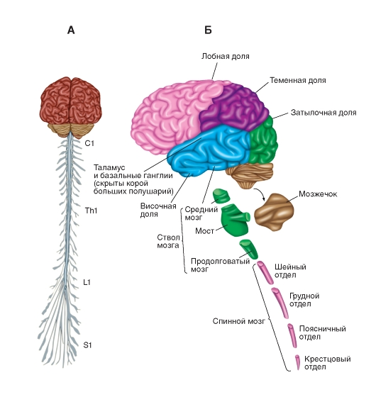
Mal. Scheme of the central nervous system (central nervous system). Dzherelo: Atlas of physiology. In two volumes. Volume 1: head. Posibnik / A. G. Kamkin, I. S. Kiselova - 2010. - 408 p. (http://vmede.org/sait/?page=7&id=Fiziologiya_atlas_kamakin_2010&menu=Fiz...)
Video: Central nervous system
The nervous system in functional and structural modifications is divided into peripheralі central nervous system (CNS).
The central nervous system is composed of headі dorsal brain.
The cephalon is located in the middle of the medulla of the skull, and the spinal cord is at the spinal canal.
The peripheral part of the nervous system is composed of nerves, tobto. bunches of nerve fibers, which extend beyond the interbrain of the brain and spinal cord and straight up to the various organs of the body. Before her, one can also bring nerve knots, otherwise ganglia- Supchennya nerve cells posture of the spinal cord and brain.
The nervous system functions as a single whole.
Functions of the nervous system:
1) shaping the awakening;
2) transmission of alertness;
3) galmuvannya (pripinennya zbudzhennya, izmenshennya yogo іtensіvі, prinіchennya, zamezhennya widening zbudzhennya);
4) integration (unification of various flows of arousal and change of these flows);
5) stimulating teasing from the outer and inner environment of the body for the help of special nerve cells - receptors;
6) coding, tobto. transformation of chemical, physical teasing on nerve impulses;
7) trophic, chi live, function - the establishment of biologically active speeches (BAS).
Neuron

Appreciate understanding
Neuron is the main structural and functional unit of the nervous system.
Neuron
- tse spetsіalіzovana vіdrostkova kіtina, zdatna spriymati, carry out that transmission of nerve impulses for processing information in the nervous system. © 2016 Sazonov V.F.
Neuron - tse folded awake secreting highly differentiated nervous clitina with teenagers as a spriymaє nerve zbudzhennya, rework yoga and pass it on to other clients. A cream of a stimulating injection of a neuron can be repaired on its own cells-targets, as well as a galvanic or a modulating injection.
The work of the galvanic synapse
Galvanic synapse on its postsynaptic membrane maє receptors to the galmic mediator - gamma-aminobutyric acid (GABA or GABA). On the surface of the excitatory synapse at the halmic synapse on the postsynaptic membrane, GABA opens ion channels not for sodium, but for chlorine. They bring to chlorine not a positive charge, but a negative one; neutralize the positive charge of sodium ions, thus stimulating the cells.
Video:The work of the GABA receptor and the halmic synapse
Ozhe, zbudzhennya through the synapse is transmitted by a chemical path for the help of special slurring speeches,what is found in the synaptic bulbs that are spread in the presynaptic plaque. Zagalna called these speeches - neurotransmitters
, then. "Neurotransmitters". Їх pod_lyayat onmediators (intermediaries), yakі transmit zbudzhennya or galmuvannya, that modulators yakі zminyuyut camp postsynaptic neuron, but zbudzhennya or galvanization themselves do not transmit.
We organize a set of clients who specialize in conducting electrical signals.
The nervous system is composed of neurons and glial cells. The function of neurons influences the coordination of functions for additional chemical and electrical signals that are sent from one month to another in the body. Most bugatoclitous creatures have nervous systems with similar basic characteristics.
Zmist:
Nervous system ingests stimuli dovkilla(External stimuli), or signals from the same organism (internal stimuli), process information and generate different reactions in the fallow situation. Like a butt, we can look at the creature, like through the cells, sensitive to the light of the network, catching the proximity of another living nature. This information is transmitted by the brain nerve to the brain, which processes and vibrates the nerve signal, and calls out the speed of the singing m'yazyv through the rukhovi nerve, so that it collapses straight ahead, to the potential problem.
Functions of the nervous system
The nervous system of a person controls and regulates the greater functions of the body, from subdivisions through sensory receptors to motor functions.
It is made up of two main parts: the central nervous system (CNS) and the peripheral nervous system (PNS). The CNS develops from the brain and the spinal cord.
NDP is made up of nerves, yakі z'ednuyut CNS z dermal part of the body. Nerves that transmit signals from the brain are called ruchial or afferent nerves, and nerves that transmit information from the body to the CNS are called sensory or afferent.
On the clitinal level, the nervous system appears to be of the clitinal type, which is called a neuron, also known as the “nervous clitina”. Neurons create special structures that allow them to quickly and accurately correct signals to other cells.
Connections between neurons can establish lances and neuron networks, thus generating spontaneity and behavior. A number of neurons of the nervous system are replaced by other specialized clitins, which are called glial clitins (or simply glia). The stench will provide structural and metabolic support.
Nervous system malfunction can be blamed on genetic defects, physical deprivation, trauma, toxicity, infection, or simply old fashioned.
Structure of the nervous system
Nervous system (NS) and two well differentiated subsystems, from one side of the central nervous system, from the other - the peripheral nervous system.
Video: Human nervous system. Intro: basic understanding, warehouse and budova

On a functional level, the peripheral nervous system (PNS) and the somatic nervous system (SNS) differentiate in the peripheral nervous system. SNR takes part in the automatic regulation of internal organs. The NDP opposes for the hoarding of sensory information and it caused voluntary changes, such as hand-drawn abo leaf.
The peripheral nervous system consists mainly of advancing structures: ganglia and cranial nerves.
autonomic nervous system
 autonomic nervous system
 The autonomic nervous system (ANS) is divided into sympathetic and parasympathetic systems. ANS takes part in the automatic regulation of internal organs.
The autonomic nervous system together with the neuroendocrine system is responsible for regulating the internal balance of our body, lowering and increasing the level of hormones, and activating the internal organs too.
For this purpose, it transmits information from the internal organs to the central nervous system through the afferent pathways and the visualization of information from the central nervous system to the m'yazyv.
It includes heart musculature, smooth skin (as it protects hair follicles), smoothness of the eyes (as it regulates the contraction and expansion of the eyes), smoothness of the blood vessels and smoothness of the walls of the internal organs (ductal-intestinal system, liver, pіdshlunkova system, respiratory system ... ).
Efferent fibers are organized, satisfying two different systems, which are called sympathetic and parasympathetic systems.
nice nervous system mainly responsible for those that prepare us to act, if we consider a significant stimulus, activating one of the automatic reactions (for example, tick chi attack).
Parasympathetic nervous system, with my own heart, supporting the optimal activation of the inner state. Increased or changed activation for the needs.
somatic nervous system

The somatic nervous system is responsible for the accumulation of sensory information. For this reason, vikoristovuyu sensory sensors, distributed throughout the body, like distributing information in the central nervous system and in this way transferring the type of central nervous system to the m'yazi that organ.
On the other side, part of the peripheral nervous system, but with voluntary control of bodily disturbances. It is composed of afferent chi of the sensory nerves, afferent chi of the rukhovoi nerves.
The afferent nerves are responsible for the transmission of intelligence to the body of the central nervous system (CNS). The efferent nerves act to increase the strength of signals from the central nervous system on the body, stimulating the contraction of the mind.
The somatic nervous system consists of two parts:
- Spinal nerves: they appear to the spinal cord and are composed of two nerves: the sensitive afferent and the other afferent dvigun, that is, the nerves are different.
- Cranial nerves: send sensory information from the neck and head to the central nervous system.
Potim resentment explained:
cranial nervous system

Є 12 pairs of cranial nerves, which are blamed on the brain and are responsible for the transmission of sensory information, control over certain diseases, and regulation of certain diseases and internal organs.
I. Olfactory nerve. We take away the scent sensory information and transfer it to the scent cibulin, which is sown in the brain.
II. optic nerve. We take visual sensory information and transmit it to brain centers zora through the sororian nerve, passing through the chiasm.
III. Internal ocular motor nerve. Vіn vіdpovіdaє for the control of the ruhіv of the eyes and the regulation of the dilatation and the shortness of the chin.
IV Intratriol nerve. Vіn vіdpovіdaє for the control of ruhіv eyes.
V. Trigeminal nerve. Vіn otrimuє somatosensory information (for example, heat, bіl, texture ...) in the form of sensory receptors in appearance and control of m'yazi chewing.
VI. External motor nerve of the ophthalmic nerve. Ruhіv eyes control.
VII. Facial nerve. We take away information about the taste of movement (things that are spread in the middle and front parts) and somatosensory information about the smell, that control of the mind, which is necessary for mimicry.
VIII. Vestibulocochlear nerve. We take auditory information and control the balance.
IX. Glossopharyngeal nerve. We take away information about the taste from the very back part of the language, somatosensory information about the language, the tonsils, the pharynx, and the control of the m'iazi necessary for forging (forging).
H. Vagus nerve. Otrimu konfіdentsіynu іnformatsiyu vіd vіd zazlechennya etching frоm tsevyih skorochenі і nadsilаєі іnformаіu іnformatsiyu і м'язам.
XI. Spinal accessory nerve. Kruє m'yazami shiї that head, yakі vykorivayutsya for ruhu.
XII. Hypoglossal nerve. Controlling the m'yazi of the tongue.

The spinal nerves feed on the organs of the mucus of the spinal cord. Nerves are responsible for the transmission of information about sensory and visceral organs to the brain and transmit punishments from the cystic brain to skeletal and smooth muscles and ridges.
The numbers are marked with reflex actions, they appear so swiftly and unfamiliarly, fragments of information can be processed by the brain to the point of being vidpovid, they are without middle control by the brain.
Usyi has 31 pairs of spinal nerves, which appear bilaterally from the cystic brain through the space between the ridges, called internal spinal openings.
Central nervous system
The central nervous system is composed of the brain and the spinal cord.
On the neuroanatomical level of the CNS, two types of speech can be seen: white and gray. The white speech is made by axons of neurons and structural material, and the speech is made by the neuron soma, decomposing the genetic material.
Tsya vіdminnіst є odnієyu z pіdstav, on some foundations of myth, in which we vikoristovuєmo less than 10% of our brain, the shards of the brain are composed of approximately 90% of white speech and only 10% of gray speech.
Ale, if you want to use speech, maybe it’s made up of material, which only serve for the purpose, today it seems that the number of such a method, for the help of which, the half-hearted vibrate, commemorately pour into the functions of the brain, shards, as if the structure is ideally located in the same place there is no sound from them, the stench is correct.
The brain is composed of impersonal structures: measles of the brain, basal ganglia, limbic system, proximal brain, stovbur and brain.

Cerebral cortex
The cerebral cortex can be divided anatomically into parts, divided by boron. The most recognizable are foreheads, tim'yans, timchasovs and potils, although the authors confirm that this is also a limbic part.
The bark is divided into two pimples, right and left, so that the halves are present symmetrically in both pimples, with the right frontal part and the left part, the right and left thyme part, etc.
The pivculi of the cerebellum are divided by an intervening fissure, and the parts are separated by various grooves.
The cerebral cortex can also be referred to the functions of sensory measles, measles of association and frontal lobes.
The sensory cortex takes in sensory information from the thalamus, as it takes in information through sensory receptors, but the primary scent measles, as it takes in information without mediation from sensory receptors.
Somatosensory information is available to the primary somatosensory measles, spread in the thymus part (in the postcentral section).
Skin sensory information reaches the main point of measles, which makes the senses of the homunculus.

As you can see, the areas of the brain, corresponding to the organs, do not follow the same order, in which the stench is spread in the body and the stench cannot be proportionately distributed.
By the largest Kirk regions, it is divided by organ sizes, that hands are ruined, shards in your eyes may be high in sensory receptors.
Visual information is available for the primary zorovoi measles of the brain, spread in the sweaty part (near the boron), and this information may be retinotopic organization.
The primary auditory cortex is found in the skeletal part (Brodmann's area 41), it is responsible for the removal of auditory information and the formation of the tonotopic organization.
The primary bark of the relish is ruffled in the anterior part of the kilchatka and the anterior tunic, and the scent bark is ruffled in the bark of the piriforms.
The cortex of association includes the first and second. The primary Kirk association is based on the sensory cortex and consequently all the characteristics of sensory information that are received, such as color, shape, appearance, size, etc. of the visual stimulus.
The root of the secondary association is found in the tim'yanіy krishechtsі and shaping the integration of information, schob praviti її to more "protruding" structures, such as frontal parts. Cі structures vm_shchuyt її in the context, to give їy sense and shy її svіdomoyu.
Foreheads, as we already guessed, are recognized for the processing of information of a high level and the integration of sensory information with ruhovymi, as they are wound in such a way that the stench responds to the stimuli that they accept.
In addition, the stench chimes a number of folding ones, ring out human tasks, as they are called vykonavchim functions.
Basal ganglia

The basal ganglion (a type of the walnut ganglion, "conglomerate", "vuzol", "puhlina") or a basal nucleus - a group of nuclei or a mass of speech (a cluster of bodies or neuron clitins), which are the basis of the brain between the upper and lower pathways of the speech tops on the brain stem.
These structures are connected one by one and at once from the cerebral cortex and association through the thalamus, their main function is to control quite a few ruhi.

The limbic system is made up of podkirkovy structures, that is, lower than the cerebral cortex. Amygdala is seen in the middle of the subkirk structures, as if to shrug, and the hippocampus is seen in the middle of the cortical ones.
Amygdala may have an amygdala-like shape and is folded from low nuclei, so that it releases afferents and visnovki from different regions.

Tsya structure is connected with a number of functions, such as emotional processing (especially negative emotions) and її influencing the process of learning that memory, respecting and deyakі mehanіzmi priynyattya.
The hippocampus, or the hippocampal illumination, is a cortical area, I look like a marine kovzan (the name of the hippocampus in the Greek hypos: a rock and a monster of the sea) and is seen in two straight lines with the brain measles and hypothalamus.
 Hypothalamus
 Tsya structure is especially important for learning, oskіlki won vіdpovіdaє for consolidating the memory, so the transformation of short-line or non-intermediate memory into long-line memory.
Intermediate brain

Intermediate brain expansions in the central part of the brain and are formed mainly from the thalamus and hypothalamus.
thalamus It is composed of decal nuclei with differentiated links, which is even more important when processing sensory information, oscills of veins coordinate that regulation information that comes from the spinal cord, the stovbur of that very brain.
In this way, all sensory information passes through the thalamus to reach the sensory measles (with a touch of scent information).
Hypothalamus folded from a large number of nuclei, like poov'yazan among themselves. In addition to other structures, both the central nervous system and the peripheral one, such as the cortex, spinal cord, network and endocrine system.
Its main function is to integrate sensory information with other types of information, for example, emotional, motivational information of the past.

Stovbur to the brain of lacerations between the middle brain and the spinal cord. Vin develops from a doubly brain, swelling and mesencephalin.
Tsya structure occupies most of the peripheral motor and sensory information, and the main function is involved in the integration of sensory and motor information.
Cerebellum
The brain is located in the back of the skull and has the form of a small brain, with a bark on the surface and with a white speech in the middle.
We take and integrate information in the main measles brain. Its main functions are coordination and adaptation of movements to situations, as well as support to balance.
Spinal cornea

The dorsal brain passes from the brain into another transverse spine. Its main function is to connect the central nervous system with the SNR, for example, by taking ruhovі commands to the brain to the nerves, like an innervation of the m'yazi, so that the stench gave a motor boost.
In addition, you can initiate automatic responses, taking away even more important sensory information, such as an injection or a liver.